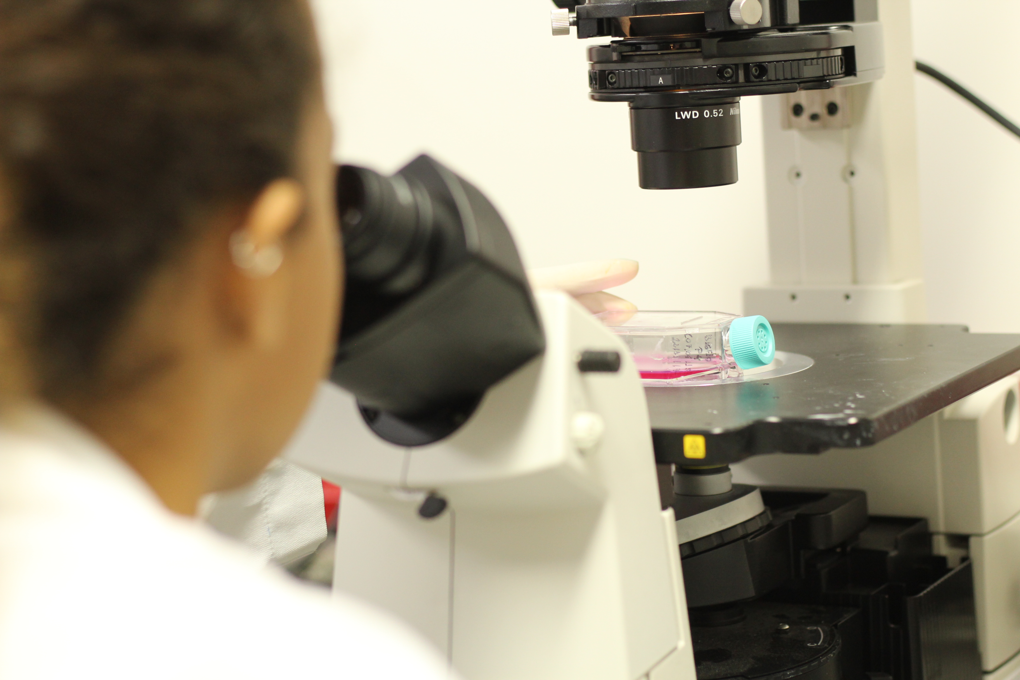
Scientists 3D-print inexpensive microscope

Scientists have 3D-printed an inexpensive and portable high-resolution microscope that could potentially be used to detect diabetes, malaria and other diseases in developing countries. The portable instrument produces 3D images with twice the resolution of traditional digital holographic microscopy, which is typically performed on an optical table in a laboratory.
It is small enough to be used in the field or at the bedside and has the potential for biomedical applications such as cell identification and disease diagnosis, according to the study published in the journal Optics Letters. In addition to biomedical applications, it could also be useful for research, manufacturing, defence and education.
"This new microscope doesn't require any special staining or labels and could help increase access to low-cost medical diagnostic testing," said Bahram Javidi from the University of Connecticut in the US. "This would be especially beneficial in developing parts of the world where there is limited access to health care and few high-tech diagnostic facilities," Javidi said in a statement.
"The entire system consists of 3D printed parts and commonly found optical components, making it inexpensive and easy to replicate," said Javidi. "Alternative laser sources and image sensors would further reduce the cost, and we estimate a single unit could be reproduced for several hundred dollars. Mass production of the unit would also substantially reduce the cost," he said.
In traditional digital holographic microscopy, a digital camera records a hologram produced from interference between a reference light wave and light coming from the sample. A computer then converts this hologram into a 3D image of the sample.
Although this microscopy approach is useful for studying cells without any labels or dyes, it typically requires a complex optical setup and a stable environment free of vibrations and temperature fluctuations that can introduce noise in the measurements. For this reason, digital holographic microscopes are generally only found in laboratories.
The researchers were able to boost the resolution of digital holographic microscopy beyond what is possible with uniform illumination by combining it with a super-resolution technique known as structured illumination microscopy. They did this by generating a structured light pattern using a clear compact disc.
The researchers evaluated the system performance by recording images of a resolution chart and then using an algorithm to reconstruct high-resolution images. This showed that the new microscopy system could resolve features as small as 0.775 microns, double the resolution of traditional systems.
Using a light source with shorter wavelengths would improve the resolution even more. Additional experiments showed that the system was stable enough to analyse fluctuations in biological cells over time, which need to be measured on the scale of a few tens of nanometres.
The researchers then demonstrated the applicability of the device for biological imaging by acquiring a high-resolution image of green algae. "Our design provides a highly-stable system with high-resolution. This is very important for examining subcellular structures and dynamics, which can have remarkably small details and fluctuations," said Javidi.
(With inputs from agencies.)
- READ MORE ON:
- Two Countries
- Nordic countries
- We Are Scientists
- Union of Concerned Scientists
- United Kingdom
- United States
- United Airlines
- Flight Facilities
- Australian immigration detention facilities
- Analog Devices
- Rhetorical device
- Output device
- countries
- Scientists
- Bahram Javidi
- computer
- microscope
- field
- world
- researchers
ALSO READ
Fans Unite: St. Pauli Raises Millions for Stadium Ownership
Crackdown at the Border: Police Forces Unite to Bust Cow-Slaughter Gang
Opposition Unites Against Controversial Waqf Amendment Bill
Shah Rukh Khan Unites Cricket and Cinema at IPL 2025 Opener
Delhi to Celebrate Odisha Parv: A Tribute to Unity in Diversity


